
Hornworms
The Anatomy of the Hornworm: A Scientific Look

A Completely Different Body Plan From a Roach
A hornworm — the caterpillar stage of the sphinx moth Manduca sexta (or the closely related M. quinquemaculata) — and a discoid roach are both insects sold as feeders on this site, and both are built from the same basic insect blueprint: a segmented body, an exoskeleton, a tracheal respiratory system. Beyond that shared foundation, the two are anatomically about as different as two insects get. Where a roach is armored in a rigid, fully hardened cuticle from head to tail (covered in detail in this site's companion piece on discoid roach anatomy), a hornworm is a soft-bodied, cylindrical animal — hardened cuticle only where it genuinely needs to be, and stretchable, flexible skin everywhere else. That single difference explains most of what follows.
The Cuticle: Hard Only Where It Has To Be
A hornworm's exoskeleton is only sclerotized (hardened) at specific points: the head capsule, the mandibles, the spiracles, the three pairs of true legs on the thorax, and the small hooked structures at the tips of the abdominal prolegs. Everywhere else — the long cylindrical trunk of the body — the cuticle stays soft and highly extensible, capable of stretching substantially between molts as the animal feeds and grows. This is a fundamentally different structural strategy than a roach's fully plated exoskeleton, and it's a direct consequence of what each animal's body needs to do: a roach needs rigid, protective armor and legs built for running, while a caterpillar needs a body that can expand dramatically to accommodate enormous, rapid feeding-driven growth, and doesn't rely on speed or armor for survival the way a roach does.
That soft-bodied structure is also part of why hornworm growth looks so dramatic to a keeper watching a cup over a few days — the stretchable cuticle allows real-time size increase between molts, not just the step-change growth spurts that happen at each molt, so a hornworm can visibly grow within a single day in a way a hard-shelled insect generally can't.
Legs: True Legs and Prolegs
A hornworm has two structurally distinct kinds of "legs," and the difference matters for understanding how the animal actually moves. On the thorax — the true insect legs, homologous to a roach's or any other insect's six thoracic legs — are three pairs of small, jointed, hardened legs near the head, used mostly for gripping and manipulating food during feeding rather than for locomotion.
The legs doing most of the actual walking are a separate structure found only in caterpillars and a few other larval insect groups: prolegs, soft fleshy stubs along several of the abdominal segments, ending in a ring or partial ring of tiny hook-like structures called crochets. Unlike the jointed true legs, prolegs work more like a hydraulic gripping system — internal muscles change the shape and pressure of the soft proleg to extend it, plant the crochets into a surface, and pull the body forward, then release and repeat down the length of the body in the wave-like crawling motion characteristic of caterpillars. This is a completely different locomotion mechanism than the claw-and-cursorial-leg running gait covered in this site's roach anatomy piece — slower, but well suited to gripping plant stems and leaves (or the smooth-ish surface of a feeder cup, which crochets can still gain some purchase on given enough surface texture) rather than to speed.
The Horn: A Defensive Bluff, Not a Weapon
The single most recognizable feature of a hornworm — the structure that gives the whole animal its common name — is the fleshy, slightly backward-curving horn projecting from the final abdominal segment. In mature larvae it's often a contrasting orange, yellow, or red against the caterpillar's green body, making it visually the most conspicuous part of the animal. Despite the intimidating look, the horn has no sting, no venom, and no piercing function — it's a soft, non-retractable projection, not a functional weapon, and handling one poses no injury risk to a person or a reptile eating one. Its actual role is believed to be primarily a visual deterrent, a bluff aimed at predators that might be put off by what looks like a stinger, alongside the caterpillar's other startle-response behaviors covered in this site's companion piece on hornworm behavior.
Spiracles and the Nicotine Connection
Running along both sides of the body are the spiracles — the same class of respiratory openings covered in this site's discoid roach anatomy piece, functioning the same basic way here: paired openings connecting to an internal tracheal network that delivers oxygen directly to tissues. In a hornworm, the spiracles are visually distinctive enough that they're sometimes described as looking like a row of small "eye" markings running down each side of the body, which occasionally confuses first-time observers into thinking they're looking at actual eyes rather than breathing structures (the animal's real, much smaller visual sensors are on the head, covered below).
These spiracles are also the specific anatomical structure at the center of this species' well-documented nicotine physiology, covered in more depth in this site's companion piece on hornworm nicotine safety: when a wild M. sexta caterpillar feeds on nicotine-containing tobacco foliage, a small fraction of that nicotine is actively transported into the hemolymph and exhaled outward through these same spiracles as a defensive chemical signal against spiders. It's a striking example of a basic respiratory structure being repurposed for an entirely different biological function — one more reason this species has become such a heavily studied model organism in insect physiology research.
Body Segmentation and the Ringed Surface Texture
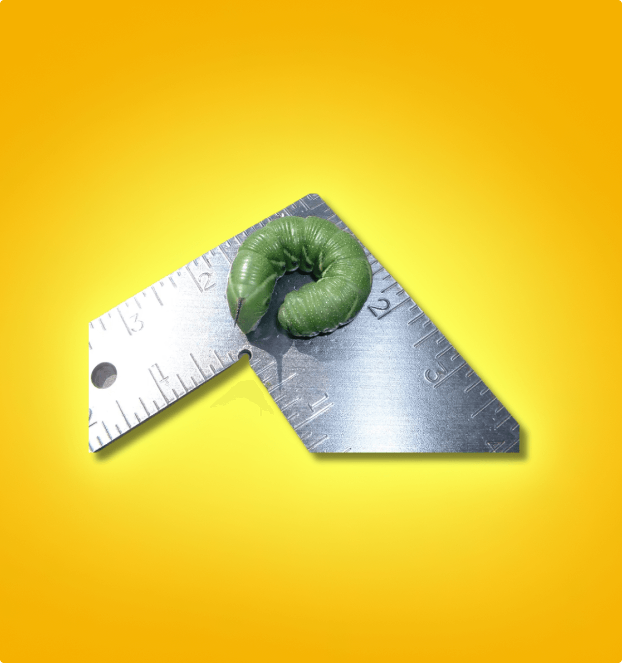
Look closely at a hornworm's body and the surface isn't smooth — each of the larva's segments is subdivided into several finer ridges called annulets, typically six to eight per segment, giving the caterpillar its distinctive ringed, almost corrugated surface texture. This subdivision isn't just cosmetic; it's part of what allows the soft cuticle covered above to flex and extend as dramatically as it does, similar in principle to how the folds in an accordion or a section of flexible hose allow expansion along a single axis without tearing the material. Combined with fine hair-like setae scattered across the body (more pronounced in later instars), the overall surface is built for exactly what a soft-bodied, rapidly growing, plant-gripping animal needs: flexibility first, protection second.
The Head: Simple Eyes and Chewing Mouthparts
Unlike an adult insect's compound eyes (covered in this site's discoid roach anatomy piece), a caterpillar's visual system is comparatively minimal: instead of compound eyes, larvae have a small cluster of simple eyespots called stemmata on each side of the head, capable of detecting light, dark, and very basic movement, but nowhere near the resolving power of an adult compound eye. This tracks with a caterpillar's actual sensory priorities — a slow-moving, leaf-bound feeding machine relies far more on touch and chemical (taste/smell) cues to locate and evaluate food than on vision, which is a secondary sense at this life stage.
The mouthparts, by contrast, are a highly capable chewing apparatus: strong, hardened mandibles built for one job — consuming large volumes of plant material as efficiently as possible to fuel the caterpillar's extremely rapid growth. This is a body built around a single overriding priority: eat continuously, grow fast, and reach pupation size before running out of time or resources — a very different life strategy than an adult insect balancing feeding against reproduction, defense, and dispersal simultaneously.
Internal Anatomy: Built for Growth, Not Longevity
Internally, a hornworm shares the same basic insect blueprint covered in this site's discoid roach piece — an open circulatory system with hemolymph moved by a simple dorsal heart, a tracheal respiratory network fed by the spiracles, and a segmented, ganglion-based nervous system — but proportioned very differently. A caterpillar's digestive tract occupies an unusually large share of the body cavity relative to an adult insect, reflecting its singular biological job: process an enormous volume of plant material relative to body size in a short window of time. Fat body tissue — the insect equivalent of a combined liver-and-fat-storage organ, present in some form across most insects — is also unusually prominent in a late-instar caterpillar, since those stored reserves are what fuel the entire non-feeding pupal stage that follows, when the animal isn't eating at all.
This is the anatomical basis for a genuinely important fact this site's practical care content already reflects: once a hornworm approaches full size and begins showing pre-pupation behavior, its priorities have already shifted from active growth to preparing for metamorphosis, and its usable window as an active, actively-growing feeder is closing.
Holometabolous Development: The Anatomy Behind Total Transformation
The single biggest anatomical fact separating a hornworm from a discoid roach isn't any one structure — it's developmental category. Roaches are hemimetabolous insects: a nymph hatches looking like a small, wingless version of the adult and gradually develops adult features (wings, reproductive maturity) across successive molts, with every instar recognizably the same basic body plan. Hornworms are holometabolous: complete metamorphosis, where the larval body plan (soft, legged, chewing, wingless) is discarded almost entirely and rebuilt from the inside out during a pupal stage into an anatomically unrecognizable adult sphinx moth — compound eyes, functional wings, a coiled feeding proboscis instead of chewing mandibles, none of which exist in any recognizable form in the larval body.
The biological machinery that makes this possible — clusters of undifferentiated cells called imaginal discs, present in the larva from a very early stage but held inactive until pupation triggers them to grow into the adult moth's eyes, wings, legs, and other structures — is one of the more remarkable pieces of insect developmental biology, and it's the direct anatomical reason a caterpillar and its own adult moth share almost no recognizable external structures in common, unlike a roach nymph and adult, which look like smaller and larger versions of essentially the same animal throughout their entire life cycle.