
Discoid Roaches
The Anatomy of the Discoid Roach: A Scientific Look

What You're Actually Looking At
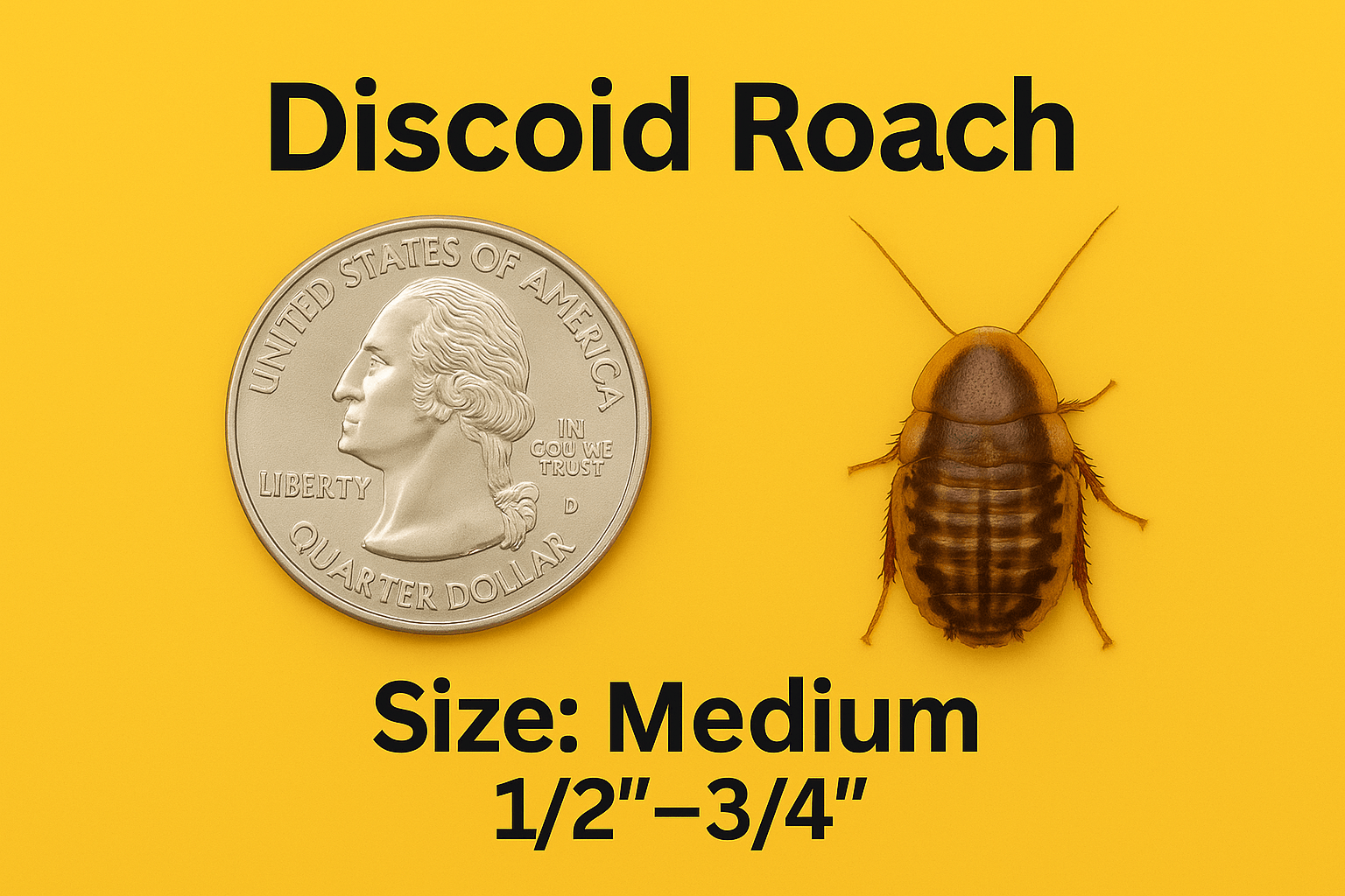
Blaberus discoidalis — the discoid roach — belongs to the order Blattodea and the family Blaberidae, the same family that includes some of the largest and most commonly kept feeder and pet roach species in the hobby (Madagascar hissing roaches and death's head roaches among them). An adult discoid roach runs roughly 35–45mm in length, tan to light brown across most of the body, with a distinctive dark brown-to-black patch across the pronotum — the shield-like plate covering the first thoracic segment, just behind the head. That patch is the single fastest visual identifier of the species and the reason "discoid" describes both the roach's flattened, oval outline and the dark disc marking its shoulders.
What follows is a systems-level walk through that body: the external plan (head, thorax, abdomen), the internal machinery that keeps it running, and the reproductive anatomy that makes this species behave differently from most of the roaches people picture when they hear the word "cockroach." None of this is trivia for its own sake — every section below connects back to something that actually matters for keeping the species, because insect anatomy and insect husbandry are the same subject looked at from two different distances.
The Exoskeleton: Armor That Doubles as Skeleton
Insects don't have an internal skeleton the way vertebrates do. Structural support comes from the outside in — a rigid but segmented external shell called the exoskeleton, built from overlapping hardened plates known as sclerites, connected by flexible membranous joints that allow movement between them. In discoid roaches, as in most Blattodea, this cuticle is coated in a thin waxy layer that serves two purposes at once: it makes the surface water-resistant, which is a real advantage for an insect that needs to retain moisture in a warm climate, and it gives the body a smooth, low-friction outer surface.
That waxy smoothness is directly responsible for one of this species' most useful husbandry traits. The muscles that would let an insect grip and climb a vertical surface need something to anchor to — either sharp claws that can dig into a rough or porous surface, or an adhesive structure that can stick to a smooth one. Discoid roaches have functional tarsal claws (more on those below) but no adhesive pad, and their cuticle's wax coating actively works against whatever marginal grip the claws might otherwise find on a glass or polished-plastic surface. The result is an animal that is structurally unable to scale the inside of a smooth-walled bin — not trained, not behaviorally reluctant, but mechanically incapable, because the anatomy required for that kind of climbing simply isn't present.
The exoskeleton is also why molting is such a significant event in a roach's life. Because the rigid cuticle can't expand, growth can only happen in bursts — the old exoskeleton splits and is shed (a process called ecdysis), and the animal is briefly soft, pale, and vulnerable until the new, larger cuticle underneath hardens and darkens. A freshly molted discoid roach looks almost white and can be mistaken for a sick or dying animal by a new keeper; it's neither. It's simply between shells, and hardening back to its normal tan coloration typically takes several hours.
The Head: Sensory Command Center
The head carries the roach's primary sensory hardware: a pair of compound eyes and a pair of long, filamentous antennae, along with the mouthparts.
Cockroach compound eyes are built from thousands of individual optical units called ommatidia — a design extensively studied in related roach species like Periplaneta americana, where researchers have documented roughly 2,000 ommatidia per eye arranged around a curved surface, each with its own biconvex lens. This produces what's called mosaic vision: rather than one sharp image the way a vertebrate eye forms one, the compound eye assembles a coarse, low-resolution composite from thousands of narrow-angle inputs. What that vision system trades away in fine detail, it makes up for in two things roaches actually need — a very wide field of view with limited need to turn the head, and strong sensitivity to movement and changes in light, which is far more useful for a nocturnal, predator-avoiding scavenger than sharp static image resolution would be.
The antennae do a different job. They're covered in mechanoreceptors (touch-sensitive structures) and chemoreceptors (smell/taste-sensitive structures), and a roach relies on them constantly — sweeping them across surfaces ahead of it to map the immediate environment, detect food, and identify other roaches by scent before any visual contact happens. In a dark, cluttered colony bin, the antennae are doing more real navigational and food-locating work moment to moment than the eyes are.
The mouthparts are the generalized chewing (mandibulate) type standard across most insects with an omnivorous or scavenging diet: a pair of hardened mandibles for cutting and crushing, paired maxillae that manipulate food and carry sensory palps of their own, and a labium forming the lower "floor" of the mouth. This is an unspecialized toolset built for a generalist diet — decaying plant matter, fungal material, and other organic debris in the wild — which lines up with why discoid roaches take readily to a gut-loading diet of fresh produce and grains in captivity; the mouthparts aren't adapted to any one narrow food source.
The Thorax and Legs: Built to Run, Not Climb or Jump
The thorax is the three-segment midsection of the body carrying the legs and, in adults, the wings. Each of the three thoracic segments bears one pair of legs, for six legs total — the defining trait of the class Insecta.
Roach legs are described anatomically as cursorial: built for running, not for jumping (like a grasshopper's enlarged hind legs) or for grasping prey (like a mantis's raptorial forelegs, notably a close relative of roaches in modern insect classification). Each leg ends in a tarsus — a small multi-segmented "foot" — tipped with a pair of claws. Those claws are excellent at gripping rough, textured, or porous surfaces: bark, soil, fabric, cardboard, unfinished wood, egg-crate flats. They provide essentially no purchase on a smooth vertical surface like glass, glazed ceramic, or polished plastic.
This is the actual mechanical answer to "why can't discoid roaches climb smooth surfaces," stated at the anatomical level rather than the observational one: some other roach species (several Blattella and Periplaneta species among them) additionally have an arolium — a soft, adhesive pad between the claws that works something like a insect's version of a suction cup, letting them grip smooth vertical surfaces the claws alone couldn't hold onto. Discoid roaches lack a functional arolium. Their tarsi are claws-only, which is precisely why a smooth-walled bin with no textured surface reaching the rim functions as complete containment without any lid modification needed for that purpose — the animal's leg anatomy simply doesn't include the hardware that would let it climb out.
The Wings: Present, But Not for Flying
Adult discoid roaches have two pairs of wings, following the standard Blattodea wing plan: a tougher, leathery outer pair called tegmina, which function more as a protective cover than as a flight surface, and a membranous, more typically wing-shaped inner pair folded beneath them. In many roach species this inner pair is a genuine flight wing. In discoid roaches, while the wings are structurally present and visible, they are not used for sustained flight — this is a species you will see walk, run, and occasionally flutter briefly, but not one that takes off and flies across a room the way some other feeder roach species can.
The wings are also the clearest, fastest way to visually sex an adult discoid roach. Males' wings extend the full length of the abdomen, covering it completely when folded at rest. Females' wings are slightly shorter relative to the abdomen, leaving the last abdominal segment or two visibly exposed past the wing tips. This is a genuinely reliable field method breeders use for colony management, and it's a direct anatomical difference (wing length relative to body length), not a behavioral or coloration cue that requires guesswork.
The Abdomen: Spiracles and the Cercal Nervous System
The abdomen is the largest body region, with roughly seven to nine visible segments depending on how the terminal segments are counted, each ringed by a hardened dorsal plate (tergite) and ventral plate (sternite) connected by flexible membrane — the same segmented-plate design as the rest of the exoskeleton, allowing the abdomen to expand somewhat (useful for a female carrying a developing brood, covered below).
Along the sides of the abdomen (and thorax) sit the spiracles — small paired openings into the tracheal respiratory system, roughly ten pairs distributed along the body in discoid roaches. Each spiracle is controlled by a small muscular valve that can open and close, which matters more than it might sound: keeping spiracles closed most of the time is a significant part of how an insect limits water loss through its respiratory surface, opening them primarily as needed for gas exchange rather than leaving them permanently open the way a mammal's airway effectively is.
At the very tip of the abdomen sits a pair of jointed, filament-like sensory appendages called cerci. These are wired into one of the best-studied rapid-response nervous systems in entomology: the cerci detect the faintest air movement — the pressure wave of an approaching predator's strike, a sudden puff of disturbed air — and feed that signal through giant interneurons directly to the leg muscles, triggering an escape run in a fraction of the time it would take for that signal to route through a more centralized decision-making process. This reflex circuit is a large part of why roaches in general have a reputation as maddeningly hard to catch by hand: the animal isn't reacting to what it sees, in many cases, but to the air current your hand created before you got anywhere near it.
What's Happening Inside: The Internal Systems
Three internal systems are worth understanding on their own, because each works differently from the vertebrate version most people are used to.
Respiratory system. Insects don't breathe through a mouth, nose, or lungs. Oxygen enters through the spiracles described above and travels through an internal network of branching tubes called tracheae, which subdivide into progressively finer tracheoles that deliver oxygen essentially directly to individual tissues and cells, bypassing the bloodstream entirely for gas transport. This is a fundamentally different respiratory architecture than a lung-and-bloodstream system, and it's part of why an insect's size is physically constrained — the tracheal-diffusion system doesn't scale up efficiently to large body sizes the way a pumped circulatory respiratory system does.
Circulatory system. Because oxygen transport is handled by the tracheal system, the circulatory system has a different job: distributing nutrients, hormones, and immune cells. Roaches, like other insects, have an open circulatory system — there's no closed network of arteries and veins the way vertebrates have. A simple tubular heart running along the back pumps hemolymph (the insect equivalent of blood, though it doesn't carry oxygen the way vertebrate blood does) forward through the body cavity, where it bathes the organs directly in an open space called the hemocoel rather than staying confined within vessels, before being drawn back in through small paired openings along the heart tube called ostia.
Nervous system. Rather than one centralized brain doing all the processing, insects run on a decentralized, segmented system: a small brain in the head connected by a ventral nerve cord to a series of ganglia — essentially local mini-processing centers — running the length of the body, roughly one per segment, each handling a lot of the reflexive control for its own local region. This decentralization is the actual anatomical basis for a genuinely true (if unsettling) fact about roaches broadly: because so much reflex control is handled locally by the segmental ganglia rather than requiring input from the head, a roach can survive for a period of time after losing its head, since breathing doesn't depend on the head (spiracles handle that independently) and basic leg reflexes don't require it either. What eventually ends that survival isn't lack of a head as such — it's the inability to eat or drink, and eventual desiccation or infection at the wound.
Reproductive Anatomy: Why Discoid Roaches Don't Lay Eggs the Way You Might Expect
This is the anatomical detail that most sharply separates discoid roaches from the roach species most people picture. Many roaches, including well-studied species like the American cockroach, are oviparous in the classic sense — the female produces an external egg case, the ootheca, and deposits or glues it to a surface where it incubates outside her body until hatching.
Blaberidae — the family discoid roaches belong to, along with Madagascar hissing roaches and death's head roaches — do something different, described as ovoviviparity (sometimes "false ovoviviparity" in stricter taxonomic usage, since the mechanism differs slightly from true ovoviviparity in some other animal groups, but functionally landing in the same place). The female forms an ootheca internally in the standard way, but instead of depositing it externally, she rotates it into a specialized internal brood sac (sometimes described as an internal uterus-like structure) and retracts it into her abdomen, where it remains through the full incubation period — typically in the range of 45 to 60 days depending on temperature and humidity, with each ootheca yielding roughly 20 to 40 nymphs. The female then gives birth to live, free-moving nymphs directly, rather than nymphs hatching from a case sitting exposed somewhere in the environment.
The practical upside of this reproductive anatomy is real: internal incubation protects the developing brood from the predation, desiccation, and mold/fungal risk an externally-deposited ootheca faces, which is part of why Blaberidae species as a group are considered relatively easy, resilient breeders in captivity — the most vulnerable stage of development happens inside the mother rather than exposed in the bin.
How This All Adds Up
None of the anatomy above is separable from how this species is actually kept and fed. The waxy, claw-only cuticle is why a smooth bin is complete containment. The tracheal-and-spiracle respiratory system, run independently of the head, is why the species tolerates handling and shipping stress reasonably well compared to more fragile invertebrates. The generalized chewing mouthparts are why gut-loading on a varied produce diet works so effectively — there's no specialized feeding structure narrowing what the animal can eat. And the internal, protected ootheca is a large part of why a starter colony reliably becomes a self-sustaining one without the keeper needing to manage a scattered clutch of externally-deposited egg cases.
Understanding the anatomy doesn't just satisfy curiosity about what's crawling around in a feeder bin — it's the actual mechanism behind nearly every practical husbandry fact this site publishes about the species, from why they can't escape a smooth container to why they arrive from shipping in reasonably good condition to why a colony builds itself up steadily rather than boom-and-bust. The behavior is downstream of the body plan.